Hydrocephalus is a rare neurological disease of cats.
By definition, hydrocephalus is an increase in the volume of cerebrospinal fluid (CSF). There are two forms of hydrocephalus:compensatory and obstructive. Compensatory hydrocephalus is the increase in volume of CSF that occurs in the cranial cavity to replace any loss of normal parenchyma.
Examples of compensatory hydrocephalus include the cerebral atrophy that occurs secondary to an infarct or the absence of cerebellum secondary to hypoplasia and atrophy caused by the in utero infection with the feline panleukopenia virus. The clinical signs relate to the loss of parenchyma not the hydrocephalus. Obstructive hydrocephalus may be congenital or acquired.
Congenital obstructive hydrocephalus results from a malformation that interferes with the circulation or absorption of CSF such as mesencephalic aqueductal stenosis or a failure of arachnoid villi to develop.
Clinical signs are present at or shortly after birth. A genetic basis is suspected but not proven for this form of hydrocephalus. Acquired obstructive hydrocephalus occurs when a disease process interferes with the circulation and absorption of CSF. These include tumors that obstruct the flow of CSF at the interventricular foramina, the mesencephalic aqueduct or the medullary lateral apertures. Tumors that compress the arachnoid villi at the venous sinuses interfere with CSF absorption.
Similarly, inflammatory diseases such as FIP meningoencephalitis or cryptococcal meningoencephalitis may obstruct the flow of CSF where the ventricular system is normally narrow. Both meningitis and ependymitis are common with these two diseases.
The clinical signs will include both those due to the primary disease as well as the hydrocephalus. The hydrocephalus that occurs in the meningoencephaloceles caused by the ingestion of griseofulvin during gestation or that seen as a genetic disorder in Burmese cats may involve compensation as well as obstruction. >Vite, Ch (2004) Developmental disorders. In: Braund’s Clinical Neurology in Small Animals: Localization, Diagnosis and Treatment. IVIS, Ithaca, New York, USA</ref>.[1].
Clinical signs
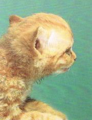
Congenital obstructive hydrocephalus is often associated with an abnormal dome shaped cranium that includes an enlarged cranial cavity and a thin calvaria with open sutures. The bony orbits are also enlarged and the eyeballs exhibit a ventroalateral strabismus probably as a consequence of the orbital malformation rather than compression of the oculomotor nerves.
The clinical neurological signs are variable but primarily relate to disturbance of the prosencephalon (forebrain) with changes in behavior, propulsive movements, blindness and seizures. More severe cases may result in signs related to compression of the brainstem and/or cerebellum. The latter will result in varying degrees of ataxia. paresis, balance loss and abnormal nystagmus. Bilateral visual defects with a normal pupillary light response is a common and consistent sign observed in affected cats. [2]. Hydrocephalus is thought to play a role in the development of hypodypsic hypernatremia in cats[3].
Diagnosis
Hydrocephalus may be confirmed by radiographic demonstration of enlarged lateral ventricles. Plain radiography will often reveal a homogenous ground glass appearance throughout the cranial cavity. Cranial sutures and/or open fontanelles may be evident after the normal age for closure and skull ossification. Ultrasonography may be performed through open fontanelles[4], CT, or MRI are the most useful diagnostic aids[5].
Collection of CSF for analysis is usually not performed since it may precipitate brain herniation due to the presence of increased intracranial pressure.
Treatment
The management of hydrocephalus depends upon establishing the cause. Many cases are not amenable to treatment.
A ventriculoperitoneal shunt has been used to treat cats with hydrocephalus[6]. Common complications include catheter blockage and sepsis.
Dexamethasone, administered at an oral dose of 1 mg/kg daily has been used empirically.
References
- ↑ Barnett, KC & Crispin, SM (2002) Feline Ophthalmology: An atlas & text. WB Saunders, London
- ↑ de Lahunta A, Cummings J. (1965) The clinical and electroencephalographic features of hydrocephalus in three dogs. J Am Vet Med Assoc 146:954-964
- ↑ Dow SW, Fettman MJ, LeCouteur RA, et al (1987) Hypodipsic hypernatremia and associated myopathy in a hydrocephalic cat with transient hypopituitarism. J Am Vet Med Assoc 191:217-221
- ↑ Rivers WJ, Walter PA. (1992) Hydrocephalus in the dog: utility of ultrasonography as an alternate diagnostic imaging technique. J Am Anim Hosp Assoc 28:333-343
- ↑ Karkkainen M. (1995) Low- and high-field strength magnetic resonance imaging to evaluate the brain in one normal dog and two dogs with central nervous system disease. Vet Radiol Ultrasound 36:528-532
- ↑ Ori J, Yamaguchi T, Komiya M, et al (1997) Two cases of feline hydrocephalus treated by ventriculoperitoneal shunt. J Jap Vet Med Assoc 50:599-602