Research on Wolbachia has caused a paradigm shift in the understanding of etiology, pathogenicity and treatment for feline heartworm disease.
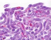
![Anti-Wolbachia spp Surface protein (WSP), showing the presence of Wolbachia bacteria within the lateral hypoderma chord of D. immitis female parasite immunohistochemistry. Courtesy August, JR (2010)[1]](https://felipedia.org/wp-content/uploads/2018/07/180px-Wolb011.jpg)
Wolbachia also facilitate parthenogenesis in insect and worms species[4], as well as evolving reproductive dependence by their host on ovum fertilisation in the presence of Wolbachia spp byproducts[5]. Wolbachia has also been found to confer on their host resistance against certain RNA virus infections[6].
Life cycle
The life cycle of Wolbachia is complex and may consist of two reproductive modes: multiplication of the bacillary forms by binary fission and by a more complex mode which resembles the Chlamydia-like cycle that consists of three morphological stages: a small, dense body, an intermediate stage with a dense inclusion, and a bacillary form which represents the final product of development and maturation of the small, dense body. The Chlamydia-like cycle offers a potential survival strategy for Wolbachia by producing more progeny than multiplication by binary fission, and appears to be more active during growth and development of embryos and of the larvae. The small, dense bodies may be the infectious forms responsible for the spread of Wolbachia through the canalicular system, within the lateral chords of filariae. An amorphous membrane that lines the perienteric surface of the body wall may represent a physical barrier that limits the spread and movement of Wolbachia to the perienteric surface of the lateral chords[7].
Wolbachia spp are believed to have an intimate relationship with most filariid parasites, including those which infect cats, especially D. immitis, Brugia malayi and B. pahangi. Wolbachia have a virtual 100% prevalence in filariids and the endosymbiontic relationship between Wolbachia spp and D. immitis is likely to be mutually beneficial insofar as Wolbachia metabolic byproducts facilitate D. immitis moulting and reproduction in the cat through the release of bacterial metabolic byproducts. Research has shown that filariid moulting in vitro is not possible in the presence of antimicrobial agents which actively suppress microbial growth[8] and that removal of the endosymbiontic Wolbachia spp from D. immitis results in their sterility and untimely death[9].
The cat can be exposed to Wolbachia when larvae, or adult D. immitis worms, are killed; when Wolbachia are expulsed, with the deposition of microfilariae, from the uterus of the females; and possibly through the excretory system of both male and female worms. The two organs that have the greatest potential of being affected by Wolbachia metabolic products/antigens released from the adult worms are the lungs and the kidneys[10].
Importance
It is now thought that Wolbachia, the bacteria which reside within D. immitis is the primary instigator of inflammation associated with feline heartworm disease[11].
It has been shown that Wolbachia is recognized specifically by IgG immunoglobulins in D. immitis-infected dogs[12] and cats[13] and that the bacteria is released into host tissue. Whether any of the clinical signs associated with feline heartworm disease are attributable to Wolbachia is not known, but it is an abundant antigen within D. immitis and its population dynamics mirror those of the filariids growth, being highest in the infective L3 to L4 stage when moulting is most active, and when inflammatory reactions by the cat against D. immitis are strongest[14].
Although Wolbachia cannot survive within mammals, their stable colonisation of filariids within mammals results in numerous pro-inflammatory effects; the bacteria trigger cytokines, neutrophil recruitment and an increase in specific immunoglobulins[15]. Thus, the logical eradication of Wolbachia as a primary therapeutic approach in feline heartworm disease seems laudable in the light of the fact that removal of Wolbachia leads to inhibition of D. immitis larval development, female worm sterilisation, and eventual adulticide of adult D. immitis.
Unfortunately, there appears to be no recorded evidence of prophylactic benefit in using antimicrobial therapy to prevent D. immitis infection, but further research may elucidate the value of this method in D. immitis-endemic areas.
References
- ↑ August, JR. Consultations in feline internal medicine. Vol 6. Elsevier Saunders, Philadelphia. pp:20-21
- ↑ Langworthy, NG et al (2000) Macrofilaricidal activity of tetracycline against the filarial nematode Onchocerca ochengi: elimination of Wolbachia precedes worm death and suggests a dependent relationship. Proc R Soc Lond B Biol Sci 267:1063
- ↑ Negri I et al (2010) Sex and stripping: The key to the intimate relationship between Wolbachia and host? Commun Integr Biol 3(2):110-115
- ↑ Knight, J (2001) Meet the Herod Bug. Nature 421:12–14
- ↑ Yen, J. H.; Barr, A. R. (1971). New hypothesis of the cause of cytoplasmic incompatibility in Culex pipiens. Nature 232: 657–658
- ↑ Teixeira, Luís; Ferreira, Álvaro; Ashburner, Michael (2008). The Bacterial Symbiont Wolbachia Induces Resistance to RNA Viral Infections in Drosophila melanogaster. PLoS Biol 6(12): e1000002
- ↑ Panteleev, DI et al (2007) The endosymbiotic bacterium Wolbachia enhances the nonspecific resistance to insect pathogens and alters behavior of Drosophila melanogaster. Genetika 43(9):1277-80
- ↑ Euclid, J (1996) Filariasis – Development of Diagnostic Assays and In Vitro Culture Studies. PhD Thesis, James Cook University
- ↑ Kramer, LH et al (2003) Immunohistochemical/immunogold detection and distribution of the endosymbiont Wolbachia of Dirofilaria immitis and Brugia pahangi using a monoclonal antiserum raised against WSP (Wolbachia surface protein). Parasitol Res 89:381
- ↑ Kozek WJ. (2005) What is new in the Wolbachia/Dirofilaria interaction? Vet Parasitol 133(2-3):127-32
- ↑ Kramer, LH (2010) The role of Wolbachia in heartworm infection. In August, JR (Ed): Consultations in feline internal medicine. Vol 6. Elsevier Saunders, Philadelphia. pp:19-26
- ↑ Kramer LH, et al (2005) Immune response to and tissue localization of the Wolbachia surface protein (WSP) in dogs with natural heartworm (Dirofilaria immitis) infection. Vet Immunol Immunopathol 106(3-4):303-8
- ↑ Morchón R et al (2004) Specific IgG antibody response against antigens of Dirofilaria immitis and its Wolbachia endosymbiont bacterium in cats with natural and experimental infections. Vet Parasitol 125(3-4):313-21
- ↑ McGarry, HF, Egerton, GL & Taylor, MJ (2004) Population dynamics of Wolbachia bacterial endosymbionts in Brugia malayi. Mol Biochem Parasitol 135:57
- ↑ Taylor, MJ et al (2001) Wolbachia bacteria in filarial immunity and disease. Parasite Immunol 23:401